Super-Resolution Microscopy
Super-resolution microscopy (SRM) describes a number of microscopy techniques that enable imaging of cellular structures at resolutions beyond the Abbe diffraction limit. This limit restricts the optical resolution in conventional light microscopy. Resolutions below the diffraction barrier reveal more details of cellular structures, which allows a better understanding of many biological processes.
Nano-Boosters, Nano-Labels, and Nano-Secondaries, which consist of Nanobodies conjugated to fluorescent dyes, penetrate tissues better and locate the fluorophore in close proximity to the cellular target because of their small size. Nano-Booster, Nano-Label, and Nano-Secondaries offers STED and/or STORM compatible fluorescent dyes:
- Alexa Fluor 488
- Alexa Fluor 568
- Alexa Fluor 647
- ATTO488
- ATTO 594
- ATTO 647N
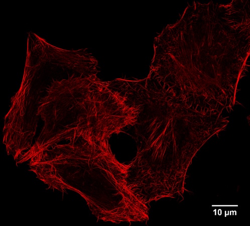
STED image of the actin cytoskeleton in HeLa cells stained with Spot-Label ATTO594.